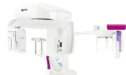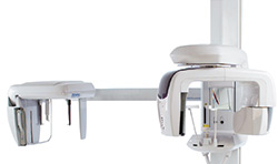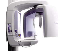
MORITA – Veraviewepocs R100/F40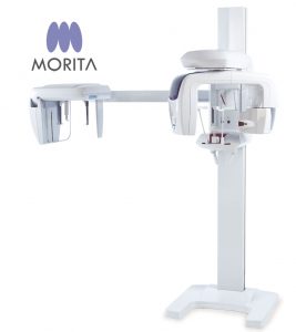

Veraviewepocs 3D R100 has changed the shape of 3D. This unit’s groundbreaking and patentet 3D Reuleaux Full Arch fields of view (FOVs) provide a unique shape for full arch imaging. With 8 field of view options and Morita’s world renowned image quality, Veraviewepocs 3D R100 is suitable for a wide variety of dental applications including implant planning.
- 3D Reuleaux Field of View
- Various Fields of View
- High Resolution Images with Dose Reduction Feature
- Easy 3D Positioning
- 3D Images for Implant Planning
- Numerous clinical cases
3D Reuleaux Field of View
Morita’s new and completely unique 3D Reuleaux Full Arch FOV abandons the typical cylinder with a new convex triangle shape. By more closely matching the natural dental arch form, this groundbreaking FOV reduces dosage by excluding areas outside the region of interest and allows a complete scan of the maxilla and/or the mandible.
Various Fields of View – Exposure Areas for Multiple Diagnostics
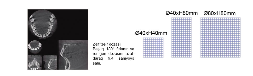
The Veraviewepocs 3D R100 model offers a total of 8 fields of view from Ø 40 x 40 mm up to Ø R 100 x 80 mm for various diagnostic needs. The new full arch scan captures the maxilla and/or the mandible with the equivalent of 100 mm in diameter and two height options of 50 or 80 mm. Its full arch capability, reduced dosage and exceptional clarity are ideal features for implant planning and oral surgery. This unit also offers small and medium field of view sizes suitable for endodontics, periodontics, as well as general dentistry.
High Resolution Images with Dose Reduction Feature
Dosage reduction program
Through advanced engineering, a Dose Reduction Mode optimizes the intensity of the X-rays which lowers exposure for easily penetrated tissues. Up to 40 % of dosage is reduced compared to the standard mode (for Ø 40 X H 80 mm exposures). By maximizing efficiency, soft tissue, such as the maxillary sinus membrane and skin, appear sharper than ever before with fewer artifacts (Compared to standard exposure mode).
Resolution & Clarity
Veraviewepocs offers high resolution images. It provides clear images of the periodontal pocket, the periodontal ligament, and the alveolar bone. It is extremely useful for implant therapy from planning to post-operative observation.
Short radiation exposure time
With a radiation exposure time of 7.4 seconds for panoramic and just 9.4 seconds for 3D images, the patient is only subjected to X-ray radiation for a brief period.
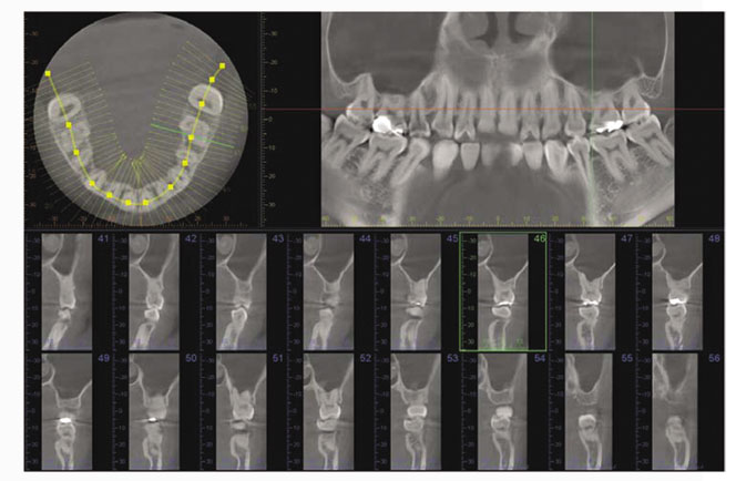
3D Images for Implant Planning
Successful placement of implants starts with the very critical and detailed planning process. Identifi cation of structures such as the sinus cavity, inferior alveolar nerve and clear views of the bone structure are needed. Veraviewepocs 3D R100 is ideal for implant planning with full arch imaging, industry leading clarity, and low dosage to the patient. This software from Morita offers advanced implant planning features, plus compatibility with popular third party software.
With this software you can highlight the mandibular canal for easier viewing, measuring the distance to the implant and determining its buccal and lingual position.